

Virginia Animal Diagnostics Newsletter - August 2021
A joint publication between Virginia Department of Agriculture and Consumer Services and the Virginia Tech Animal Laboratory Services
Editorial:
Pathology Over Time
This might also have been “Pathology Overtime” due to my status as the oldest one around now. It might help others to share a bit of a longer perspective on a regional perspective of diseases and their incidence. A few infectious diseases tend to cycle periodically. While the cycle varies from disease to disease, most follow a pattern of being evident for a year or two, then disappearing for several years, then recurring once again. In some situations, they become relatively common during those periods of increase.
One disease that has a multi-year cycle in our region is canine distemper. One signal, at least locally, is a dramatic uptick in the number of racoons that are dead on the side of the road, although a good outbreak of rabies can do the same thing! I have not kept track of the periodicity of canine distemper in dogs, but it does seem to appear for a few years, and then disappear for something on the order of five years or so. It is fairly routine, at least for me, for submitting clinicians question any diagnosis of canine distemper, because “that disease is gone.” It just so happens that canine distemper is never “gone for good.” It must always be on a list of differentials, especially for those cases in young dogs that combine respiratory signs with signs related to the central nervous system. Especially relevant in today’s human coronavirus pandemic is the need for diligence in vaccination. Adequate vaccination, and adequate handling of vaccines, are essential.
Another disease that tends to cycle over multiple years is salmonellosis in farm animals. The lab here has a minimal caseload in poultry, so this observation is more along the lines of horses, cattle, sheep, and goats. Some years see a dramatic increase in cases infected with Salmonella, and then several years will pass by with no positive cases. This can easily lull the diagnostician into a false sense of security, with the very real possibility that cases could be missed. The epidemiology of salmonellosis is important, with some outbreaks having an annoying pattern that suggests traveling along with veterinary clinicians.
A few others, such as carnivore parvovirus infections in cats and dogs, do not seem to cycle and never seem to either surge all that strongly, nor do they tend to disappear. This disease can also cause both clients and submitting clinicians to question a diagnosis because some of these cases have happened in vaccinated animals. Once again, vaccination protocols become an important potential weak link in the chain of protection. Fortunately, this disease has a characteristic pattern of lesions, so could only be confused with something like radiation poisoning. When I bring up that possibility, the conversation usually changes fairly quickly. I have yet to call the National Security Agency, and hope that it stays that way.
I am sure there are other diseases, generally infectious ones, that have cycles of occurrence, but these are the few that stand out most strongly to me.
Phillip Sponenberg DVM, PhD, Virginia Tech.
Equine and Camelids
Potomac horse fever
A deceased 25-year-old Quarter Horse gelding was submitted to the Harrisonburg RAHL with a short history of diarrhea, fever, increased heart rate and depression. Treatment with fluids and tetracycline was unrewarding and the equine’s condition deteriorated. The owners elected euthanasia. The stomach contained green ingesta with the small intestine having segmental mucosal reddening and dilatation. Dilated small intestine in the distal segment had dark red mucosa often mottled with scattered irregular gray areas. These features extended into the cecum and proximal portions of the dorsal right colon. Distal large colon mucosa was markedly edematous with watery green contents. Histopathology of the small and large intestine revealed large numbers of lymphocytes and plasma cells expanding the lamina propria, extending into and expanding the submucosa. Referral testing of EDTA collected blood was positive by polymerase chain reaction (PCR) testing for Neorickettsia risticii. Historically, Potomac Horse Fever was first noted in 1979 in the Eastern United States around the Potomac River. It was a sporadic disease in equines characterized by fever, diarrhea and other signs such as laminitis and possible abortion. Research identified the causative organism as a Neorickettsia, with intermediate stages of trematodes and aquatic insects such as damselflies, mayflies, and caddis flies as the transmitting organisms.
David Brown DVM, RAHL Harrisonburg.
Equine rhodococcosis
Rhodococcus equi pneumonia was the cause of death in a five-week-old American Saddlebred foal. The foal became acutely ill with fever (104.5F), dyspnea and an elevated WBC (14,000). Despite treatment with Clarithromycin and rifampin, the foal succumbed approximately 48 hours later. Gross pathology revealed severe bilateral pneumonia with large multifocal and coalescing abscesses. Histopathology confirmed necrotizing abscessation composed of central cores of acellular and necrotic debris, fibrin, degenerate and non-degenerate neutrophils and aggregates of bacteria. Aerobic culture of the affected lung tissue resulted in the isolation of both Rhodococcus equi and Streptococcus equi ssp. zooepidemicus. The owner reported a prior history of similar cases on this premises.
Christopher Halsey DVM, RAHL Wytheville.
Equine motor neuron disease
A five-year-old Warmblood cross mare was humanely euthanized and submitted for necropsy following a history of chronic, intermittent ataxia and weakness that was initially responsive to treatment for Equine Protozoal Myeloencephalitis, but relapsed after two treatments. No significant gross lesions were detected, but a spectrum of lesions were detected microscopically in the neuraxis including: chromatolysis and degeneration of neurons within the spinal cord ventral horn and brainstem nuclei associated with intraneuronal lipofuscin accumulation, axonal swelling in the affected gray matter, and Wallerian-type degeneration bilaterally distributed throughout all funiculi in the spinal cord. These findings were compatible with a diagnosis of equine motor neuron disease, a term that encompasses a group of neurodegenerative diseases through to result from primary metabolic dysfunction of the nerve cell body. In some cases, vitamin E deficiency appears to contribute to the pathogenesis.
Thomas Cecere DVM, PhD, DACVP, Virginia Tech.
Ruminants
Theileria orientalis
Theileria orientalis was detected in anticoagulated whole blood via PCR in a two-month-old Angus cross bull calf that was sick for one-week duration and anemic (PCV 10%). The calf died shortly after transfusion and was submitted for necropsy. Gross and microscopic examination revealed lesions of hemolytic anemia, including icterus and hepatosplenomegaly. Touch imprints of the spleen taken at necropsy revealed multiple intracellular piroplasms within erythrocytes, consistent with the diagnosis of Theileriosis.
Thomas Cecere DVM, PhD, DACVP, Virginia Tech.
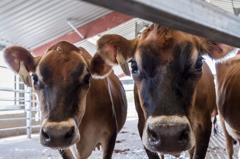
Cryptosporidosis in a calf
A ten-day old Jersey heifer was presented for necropsy following multiple days of diarrhea and subsequent spontaneous death. The distal small intestine, cecum, colon, and rectum contained abundant yellow fluid material but the intestinal mucosa and serosa were grossly normal. Laboratory findings included Cryptosporidia spp. on fecal flotation. Histologic examination revealed protozoal organisms attached to the epithelial surface of the colonic mucosa, along with neutrophils in the crypts. This is a case of cryptosporidiosis.
Phillip Sponenberg DVM, PhD, Virginia Tech.
Pigs and Birds

Listeriosis in a chicken
A chicken with history of lethargy and anorexia was submitted dead for necropsy. On gross examination, the only abnormality was that 80% of the heart was firm and white on cut surface. Microscopically, the heart lesions were consistent with granulomatous myocarditis. There was also evidence of fibrin thrombi in multiple organs and splenic necrosis suggesting a systemic bacterial infection. Bacteriology cultures revealed growth of Listeria monocytogenes in the heart and spleen. Listeria monocytogenes infection is most commonly reported in cattle, sheep, and goats where it is typically associated with neurologic disease. However, it can rarely be seen in poultry where it more commonly results in a septicemia primarily affecting the spleen, liver and heart. L. monocytogenes is a gram-positive bacterium that is commonly found in the environment, especially soil, feces, and decaying vegetation. Infection occurs following inhalation, ingestion, or wound contamination with the bacterium but chickens and turkeys are relatively resistant to infection. Contamination of poultry farms with fecal material from nearby food animal farms, especially after a rain or flooding, can contribute to outbreaks in poultry. Prevention primarily focuses on identifying and eliminating potential sources of infection, which can be difficult in some cases. There is zoonotic potential of listeria. Humans can become sick following exposure to raw or uncooked poultry products.
Jaime Weisman DVM, MS, RAHL Warrenton.
Toxoplasmosis in a piglet.
A 2-week-old piglet with history of tremors and ataxia was submitted for necropsy. No significant gross findings were identified. On histology, sections of white and grey matter of the brain were obscured by multifocal random foci of dense cellular infiltrates composed of lymphocytes, plasma cells, and macrophages, plus multifocal areas of neuronal necrosis. Sections at the level of the hypothalamus contain neurons with multiple small round to pear shaped apicomplexan organisms. This apicomplexan parasite is likely Toxoplasma gondii. This organism is relatively common in domestic species and can be transmitted by ingestion of food/water contaminated with oocysts, ingestion of rodents with tissue cysts, or likely in this case, congenital from an infected sow.
Valentina Stevenson DVM, Virginia Tech.
Companion/Exotic Animals

Lymphoma in a dog
A nine-month-old female Labrador retriever dog presented to the emergency service at the Virginia-Maryland College of Veterinary Medicine Veterinary Teaching Hospital for acute onset of dyspnea and paraplegia. Based on the ensuing clinical workup, systemic mycosis with pulmonary and CNS involvement was suspected. Humane euthanasia was elected and the dog was submitted for necropsy, which revealed multicentric lymphoma involving the mediastinum, lung, multiple visceral lymph nodes, kidney, spleen and a focal epidural mass associated with spinal cord compression.
Thomas Cecere DVM, PhD, DACVP, Virginia Tech.
Amanita intoxication
A puppy, an older dog, and a mature cat were presented from different owners all in the same week. Each had a history of rapid decline. All three had massive necrosis of the liver, which was nearly complete in most of them. This is consistent with consumption of Amanita spp mushrooms. In each of these cases further history was obtained, and each of these animals had consumed mushrooms.
Phillip Sponenberg DVM, PhD, Virginia Tech.
Laboratory News
ViTALS
ViTALS is thrilled to announce the addition of three new faculty members to the pathology group. Dr. Teresa Southard is an Associate Professor of Anatomic Pathology, and comes to us from Cornell College of Veterinary Medicine. At Cornell, she was Chief of the necropsy service, and Head of the TSE laboratory. Dr. Vanessa Oakes joined us in June as a clinical instructor in Anatomic Pathology. Dr. Oakes recently finished her pathology residency and will take the ACVP phase II exam this month. Dr. Priscilla Serpa will start in September after finishing her clinical pathology residency at Purdue University and taking phase II of her ACVP board exam. We are very excited that they have decided to join our team.
Dr. Katie Boes will be leaving Virginia Tech this month, but we are happy that she will be staying in Blacksburg. She played a critical part in developing and delivering the new DVM curriculum, and in growing the clinical pathology lab into the largest section of ViTALS. She will be missed.
We are in the process of standing up new tests for Chronic Wasting Disease testing in deer. New tests are being validated in clinical pathology, including Free T4, fibrinogen, D-dimers, and platelet counts, and will be available soon.
Office of Laboratory Service
New Serologist in Harrisonburg- Josh Yother; New Business Manager for VDACS OLS- Anne Magee, previously the dairy microbiologist in our Ivor lab! Welcome!
The Harrisonburg RAHL is excited to announce the purchase of a new, state of the art, MALDI-TOF Biotyper. This machine will assist in the rapid identification of bacterial pathogens by the use of a laser with little biochemical testing required. We are looking forward to being able to utilize this platform to rapidly identify disease-causing organisms to assist in making treatment decisions.
Laboratory Locations
HARRISONBURG
261 Mt. Clinton Pike
Harrisonburg, VA 22802
540-209-9130
RAHLHarrisonburg@vdacs.virginia.gov
WARRENTON
272 Academy Hill Rd.
Warrenton, VA 20186
540-316-6543
RAHLWarrenton@vdacs.virginia.gov
LYNCHBURG
4832 Tyreeanna Rd.
Lynchburg, VA 24504
434-200-9988
RAHLLynchburg@vdacs.virginia.gov
WYTHEVILLE
250 Cassell Rd.
Wytheville, VA 24382
276-228-5501
RAHLWytheville@vdacs.virginia.gov
Virginia Tech Animal Laboratory Services
205 Duck Pond Drive
Blacksburg, VA 24061
540-231-7666
lcrvth@vt.edu