

Virginia Animal Diagnostic Newsletter - November 2021
A joint publication between Virginia Department of Agriculture and Consumer Services and the Virginia Tech Animal Laboratory Services
Editorial:
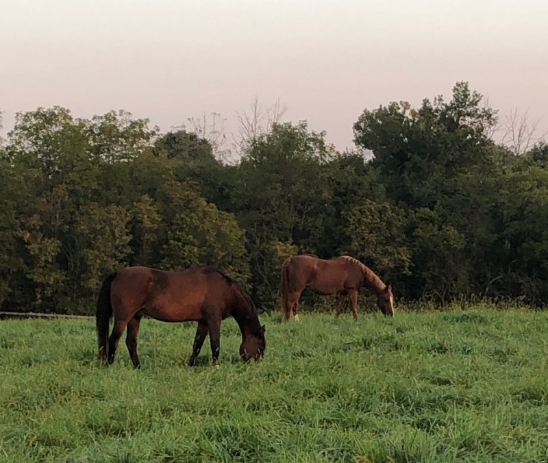
Pneumonia in horses from the pathologist's perspective: what are we missing?
I have been lucky enough to train and work under the guidance of great veterinary pathologists. My previous job at the California Animal Health and Food Safety Laboratory in San Bernardino, CA introduced me to a very interesting field of pathology: racehorse pathology. Most of the equine cases presented to the diagnostic laboratory were horses with musculoskeletal issues. The second most frequent set of cases were horses with severe pulmonary lesions.
Interestingly, most racehorses with pneumonia had been treated with different antibiotics for weeks. Some of them recovered and some of them succumbed to a severe pleuropneumonia, characterized by the presence of abundant fibrinosuppurative exudate in the thorax, sometimes with a foul smell. I could not believe that horses could survive a long time with such a severe lesion! We started looking a little closer at those cases, and we realized several facts that are believed to be true of several species of animals, but that do not apply in all their details to horses.
The paradigm taught in every veterinary school is that pneumonia in animals is a bacterial process that is frequently secondary to a primary viral infection. As veterinary students, and then as veterinary practitioners, we tend to stay within those boundaries. We try to find a viral disease behind every case. For ruminants and pigs, this is mostly true. A few other factors are also recognized in dogs and cats, such as stress and overcrowding, and these factors can also apply to large animals.
In horses, the scenario is a bit different. Several studies in racehorses have failed to link a viral infection with severe pneumonia. Other studies have found that the most important predisposing factor for pneumonia in racehorses is transportation! There are several reasons for that. First of all, horses are transported for long hours, some of them on the highway in open trailers, so that desiccation of airways plays an important role. This is due to malfunction of the mucocilliary apparatus. Second, some horses cannot move their heads during transportation, which does not allow them to clear their airways with sneezing or neck movement. Adding other stressors of transportation such as highway noise and other trucks, and the result is a perfect storm that allows bacteria in the upper airways to reach the lung. Racehorses have yet other risk factors that occur in between these periods of transportation from one racetrack to the next. While racing and training, racehorses inhale dirt from the racetrack, which certainly adds both stress and contaminated dirt to the respiratory system.
By far the most common bacterium isolated from cases of pneumonia in racehorses is Streptococcus equi ssp. zooepidemicus (S. equi zoo), alone or in combination with other aerobic and anaerobic bacteria. S equi zoo is a normal commensal bacterium of the skin, nasal cavity and oral cavity in horses. It is important to consider that when horses with pneumonia die or are euthanized and submitted for necropsy, they have been treated for several days (sometimes weeks) with different antibiotics. Several studies have sought to identify virulence factors that confer S. equi zoo an adavantage for colonization and infection in the equine lung, but it has been found that there is no single pathogenic trait that confers this bacterium advantage over other bacteria. Plus, several strains of S. equi zoo can be isolated from a single case! So, what is the meaning of this? Are we actually isolating the “smoking gun” or are we isolating the bacterium with best survival abilities, which could have invaded the lung after the primary damage was done? This is still matter of debate. Despite these uncertainties, the underlying truth remains that viral agents do not play a major role in the development of the lesion.
Our case load in Virginia is mostly non-competing horses, yet we have found that S equi zoo is also the most frequently isolated bacterium in cases of equine pneumonia within this population. This is not completely unexpected, because “backyard” animals often have their own set of stressors. Many of them are transported for various purposes and this provides the same stress for them as it does for race horses. There are certainly other factors that we are missing as mere observers of cases in the necropsy floor. This is where ventilation, cold weather, and overcrowding can each play a role. In these cases, no microscopic lesions suggestive of a viral infection are observed in the respiratory system, but we do have to consider that histopathology is not a very sensitive method for the identification of viral pathogens.
If something has to be learned from these cases in two locations with very different equine populations, it is that horses should not be transported for more than 5 hours without having the opportunity to “stretch their legs”, move their head, and sneeze. A little stop along the way for a coffee (for the driver) and a walk (for the horse) will certainly increase the chances of the horse not developing pneumonia. This is important, because pneumonia frequently has fatal consequences in horses. Regarding the infectious cause, we will likely continue dealing with S. equi zoo for long years, but we have to consider that it may not be the “smoking gun”… What else are we missing?
Francisco R Carvallo DVM, DSc, DACVP
Veterinary Anatomic Pathologist
Virginia Tech and VDACS
Fcarvallo@vt.edu
Horses
An atypical Salmonella enterica isolate in a horse
A 4-day old foal presented for treatment of diarrhea. During hospitalization, the foal’s diarrhea did not resolve and, despite antimicrobial therapy and nursing care, she developed clinical signs and hematologic abnormalities associated with sepsis. Salmonella was isolated from a blood culture. The foal continued to decline and was ultimately euthanized. Her dam remained healthy but was screened for Salmonella and her fecal culture was positive. The isolates from both the foal and her dam were determined to be Salmonella enterica serotype Hadar. Salmonella serotyping is a classification scheme used for epidemiologic and surveillance purposes that includes more than 2500 serotypes, which are based on surface antigens on the bacteria. Salmonella Hadar is an unusual serotype in horses, and has only been recently described in a horse in the United States; it has previously been described in equine cases in Europe. Salmonella Hadar is more commonly associated with poultry and foodborne illness in people. Salmonella serotypes frequently isolated from horses include Typhimurium, Newport, Muenster, and Montevideo. Serotyping can be an important part of working up salmonellosis; as in this case, we can use serotyping data to detect incursions of strains into new locations or potential links between infected animals.
Tessa LeCuyer DVM, PhD, DACVM, Virginia Tech.
Potomac horse fever
A fifteen-year-old Appaloosa mare was seen by a veterinarian for watery diarrhea and inappetence. Following initial evaluation, the horse was referred to the VMCVM Veterinary Teaching Hospital with azotemia, severe diarrhea, and dull mentation. Based on declining status, humane euthanasia was elected and the horse presented for necropsy. The small intestine, cecum, and colon were distended throughout and filled with green-tinged watery fluid. There was segmental lymphoplasmacytic typhlocolitis with marked submucosal edema, most severe within the right dorsal colon. No pathogens were detected on routine fecal culture, and Clostridium difficile toxin was not detected on ELISA. Feces submitted to the University of California Davis School of Veterinary Medicine was qPCR positive for Neorickettsia risticii, and qPCR negative for Equine Coronavirus, Clostridium difficile toxin A and B, Lawsonia intracellularis, and Salmonella spp. Neorickettsia risticii is the causative agent of Potomac Horse Fever, a syndrome of enterocolitis usually seen in spring, summer and early fall which involves a trematode vector. In addition to gastrointestinal disease (colic, diarrhea), approximately 20-30% of affected horses develop concurrent laminitis.
Thomas Cecere DVM, PhD, DACVP, Virginia Tech.
Ruminants

Bovine Respiratory Syncytial virus
A yearling Angus bull was submitted to the VDACS Wytheville Regional Lab for necropsy. The farm recently had four cases of suspected respiratory illness. This particular animal had demonstrated clinical symptoms that included fever and expiratory grunt. There was no response to medical therapy (florfenicol, flunixin). Post-mortem examination revealed gross lesions of multifocal (primarily cranio-ventral) congestion/consolidation of both lungs. Interstitial emphysema was also noted. Aerobic culture of the affected lung resulted in no bacterial growth. Histopathology revealed severe, fibrinosuppurative broncho-interstitial pneumonia with type II pneumocyte hyperplasia, rare multinucleate cells, and interlobular edema and emphysema. Lung tissue was forwarded to the Texas Veterinary Medical Diagnostic Laboratory for a Bovine Respiratory Disease PCR Panel. Results were positive for Bovine Respiratory Syncytial Virus.
Christopher D. Halsey DVM, RAHL Wytheville
Septicemia in calves
There was an outbreak of disease in a herd of calves birthed aseptically and maintained in a clean environment as part of a research project. Initial clinical signs were subtle and included fever and general malaise; bloodwork revealed lymphopenia, increased band neutrophils, and hypoproteinemia. On necropsy, the animal had a severe, diffuse fibrinous enteritis with pseudomembrane formation, fibrinous peritonitis, adrenal gland corticomedullary hemorrhage (Waterhouse-Friderichsen syndrome), and fibrinous arthritis in multiple joint spaces. Salmonella spp. and E. coli were cultured from the affected animals. The point of contamination is still under investigation.
Vanessa Oakes DVM, DACVP, Virginia Tech.
Hydrops allantois with adventitial placentation
A 5-year-old late gestation Jersey cow was presented for necropsy with a history of abdominal distension, tachycardia, scant feces, and copious uterine fluid with an inability to palpate the fetus. The uterus was filled with greater than 100 liters of straw-colored fluid that was primarily within the allantois and a single fetus. The intercotyledonary placenta contained numerous coalescing pink/tan nodules ranging in size from 0.2-1cm diameter that resembled placentomes microscopically. The necropsy findings were consistent with hydrops allantois with adventitial placentation. Adventitial placentation is the development of intercotyledonary placentation to compensate for inadequate development of placentomes, and is associated with hydrops allantois.
Thomas Cecere DVM, PhD, DACVP, Virginia Tech.
Companion Animals
Aspergillosis in a dog
A six-year-old, intact female, mixed-breed dog was presented to the hospital given a history of recurrent urinary tract infection (UTI) for 2 years. Complete blood count and serum chemistry panel were unremarkable. However, the urinalysis revealed an inadequate urine specific gravity and an active sediment. Pyuria and hematuria were present, as well as low numbers of fungal hyphae. On culture, Aspergillus terreus was identified. The treatment consisted of removing her left kidney, which was effaced by a granuloma with intralesional fungal hyphae. There was no evidence on history or clinical data that the patient was immunocompromised.
Priscila B. S. Serpa, DVM, MSc, DSc, DACVP (Clinical), Virginia Tech.
Protothecosis
A 2-year-old, female spayed husky mix submitted for necropsy for chronic hematochezia. On gross examination there was prominent cutaneous petechial hemorrhages, a myriad of pinpoint foci in the liver and kidney, and marked mucosal thickening of the cecum and colon. Impression smears of the liver, colon, and kidney revealed organisms consistent with Prototheca species. Similarly, microscopic evaluation revealed disseminated algal infection consistent with Prototheca species in multiple tissues. Prototheca are opportunistic algae found in the environment that rarely causes disease in dogs and other animals. Protothecosis is most commonly associated with gastrointestinal disease in dogs but can also result in ocular, cutaneous and disseminated disease. There is no successful known treatment for protothecosis.
Jaime Weisman DVM, MS, RAHL Warrenton.
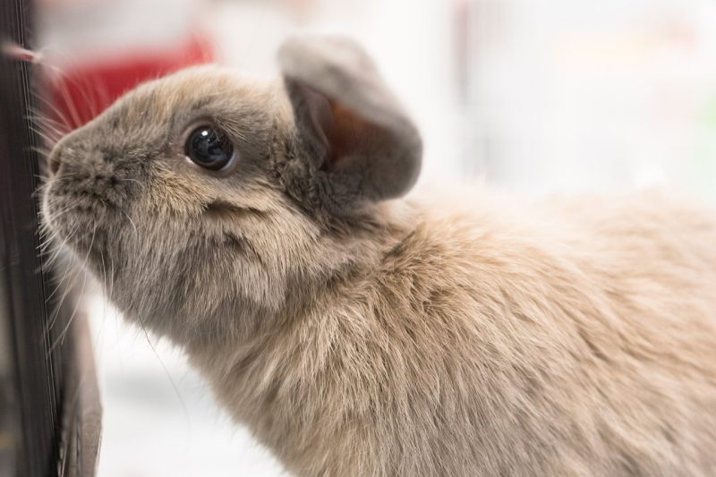
“Snuffles” in a rabbit
A 6-year-old, male neutered, Holland lop rabbit was presented to the VMCVM necropsy service following an 8-month history of sneezing and difficulty breathing. Chest radiographs revealed a soft tissue opacity mass occupying part of the thoracic cavity. On necropsy, the rabbit was found to have approximately 100 mL of opaque, red-tinged fluid in the thoracic cavity. There were multiple, coalescing abscesses completely effacing the left lung lobes, measuring up to 3cm in diameter, and containing tenacious, white, caseous material. Although there were several abscesses in the right lung lobes, they were much smaller; the remaining lung was atelectatic. These clinical findings are consistent with “snuffles”, a common disease in rabbits. The causative agent of snuffles is Pasteurella multocida. Typically, “snuffles” presents as an upper respiratory infection, but with chronicity, it can extend into the lungs, where it can cause abscessation. Otitis is a common sequela, although the ear canals were unaffected in this case. The soft tissue opacity masses seen radiographically were the multiple pulmonary masses in this case. Contributing to the patient’s dyspnea was the compression of the unaffected lung lobes, both by the space-occupying abscesses, and by the inflammatory fluid in the chest.
Vanessa Oakes DVM, DACVP, Virginia Tech.
Laboratory News
ViTALS:
- We are excited to announce that Dr. Natalia Strandberg will be joining the ViTALS team on December 16th, 2021 as a clinical instructor of Clinical Pathology. Dr. Strandberg was a clinical pathology resident at Purdue, and is a Diplomate of the American College of Veterinary Pathology. In more good news, Dr. Vanessa Oakes (Anatomic Pathology) and Dr. Priscila Serpa (Clinical Pathology) both passed the ACVP board certifying exam. Congratulations to Dr. Strandberg, Oakes, and Serpa for this huge accomplishment!!
- Theileria orientalis Ikeda has been identified in the mid-Atlantic region over the last 4 years with increasing range and prevalence. This is a tickborne disease that can lead to anemia, death and abortion in a percentage of infected cattle. In order to give more frequent updates about this disease, ongoing work, and potential collaboration, we have created a webpage. The first entry covers ongoing investigation into abortions and a request for collaboration. http://vitals.vetmed.vt.edu/theileria
VDACS:
- The VDACS Regional Laboratory System is continuing to consolidate and expand testing to better serve the needs of our clients. The online fee schedule with test prices, location, and preferred specimen type has been updated and will be reviewed regularly for any potential changes. We have been unsuccessful in hiring a Laboratory Director for the Lynchburg Laboratory, but have maintained all testing services except necropsy at that location. We will soon be offering necropsy kits for field necropsy submissions, as well as a wider range of PCR testing for diagnostic submissions. We are excited to continue to work with our clients and stakeholders to make our laboratory system even better in serving our Commonwealth!

Laboratory Locations
HARRISONBURG
261 Mt. Clinton Pike
Harrisonburg, VA 22802
540-209-9130
RAHLHarrisonburg@vdacs.virginia.gov
WARRENTON
272 Academy Hill Rd.
Warrenton, VA 20186
540-316-6543
RAHLWarrenton@vdacs.virginia.gov
LYNCHBURG
4832 Tyreeanna Rd.
Lynchburg, VA 24504
434-200-9988
RAHLLynchburg@vdacs.virginia.gov
WYTHEVILLE
250 Cassell Rd.
Wytheville, VA 24382
276-228-5501
RAHLWytheville@vdacs.virginia.gov
Virginia Tech Animal Laboratory Services
205 Duck Pond Drive
Blacksburg, VA 24061
540-231-7666
lcrvth@vt.edu