

Virginia Animal Diagnostics Newsletter - February 2021
A joint publication between the Virginia Department of Agriculture and Consumer Services and the Virginia Tech Animal Laboratory Services
Veterinary Forensics:
Handling and documentation of evidence 101
With the growing number of animal related investigations, there is an increased demand for veterinary expertise in cases of possible animal cruelty. Based on this need, veterinary forensics is emerging as a distinct discipline, but many veterinarians do not feel they have received adequate training to handle these cases. Veterinary forensics is simply the application of veterinary knowledge to answer questions of interest to a court of law.
Proper handling of evidence and documentation of that evidence is necessary in any potential forensic case. Failure to properly handle or document evidence can make or break a case. A chain of custody should be used to document anyone who comes in contact with a piece of evidence. Include the individual relinquishing the evidence, the receiver, and date. Without a proper chain of custody, evidence collected may be challenged in court and possibly considered compromised. In animal investigations, the animal and any sample taken from the animal are considered evidence and should, therefore, be tracked on a chain of custody. An evidence log is often used in conjunction with the chain of custody to provide a description and unique ID for each item of evidence. A simple internet search can be used to find many examples of chain of custodies and evidence logs that can be useful in creating such documents for veterinary practices.
Photography and/or videography are also a vital part of documentation and should be utilized in all forensic cases. In this discussion, I will focus on photography, but videography may also be applicable in some cases to better document wounds, specific behaviors, pain, and various other abnormalities. Photographs are not only important in documenting injuries and wounds but are also used to demonstrate improvement following treatment. For example, weekly photographs of an emaciated animal to demonstrate weight gain with proper nutrition can be crucial in a possible starvation case. A simple white board can assist with coordinating a series of pictures with a specific animal by starting each series of pictures with a dry-erase board containing the animal's identification information, date, and any other relevant information. This can be especially helpful when there are multiple animals involved in a case. Pictures of the animal should also include all identifying marks. This can be accomplished by taking pictures of the right and left lateral, ventral, and dorsal aspect of the animal in addition to the face and any other unique markings. When photographing lesions, it is recommended to take pictures at a distance to better determine orientation, close-up to identify small details, and with a ruler for sizing. Ideally, all pictures should be taken perpendicular to the evidence to prevent distortion that may result from oblique camera angles. Any ruler can be used for sizing purposes, but there are many photographic scales that are designed specifically for forensic documentation (e.g. ABFO No. 2 L-shaped ruler) that allows for better photographic documentation. When organizing pictures it is essential to save a non-doctored copy of each picture taken to ensure transparency of the evidence. It is also important that any picture, even those of poor quality, are not deleted. A photo log, including picture identification and description of each picture, can be helpful when reviewing a case at a later date and can assist the court in identifying important features of a picture.
The above information is only a brief introduction to handling potential forensic cases but there are many resources, including multiple publications, continuing education courses, and certification/masters programs, for those who want more advanced information in veterinary forensics.
Jaime Weisman, DVM, MSc, RAHL Warrenton
Jaime.weisman@vdacs.virginia.gov
Equine

Nocardioform placentitis
A late-term aborted horse fetus was submitted for necropsy. The fetus had minimal changes that included faint petechial hemorrhages in the lungs and congestion of multiple organs. However, the chorioallanotic portion of the placenta was covered with large amounts of thick, brown exudate. These findings are indicative nocardioform placentitis (atypical placentitis) which was confirmed on bacteriology cultures. This form of placentitis is associated with a group of similar organisms including Crossiella equi, Amycolatoopsis kentuckyensis, Amycolatopsis lexingtonensis and Amycolatopsis pretoriensis that resemble nocardia species. Nocardioform placentitis can result in a late-term abortion, stillbirth, or premature birth.
Jaime Weisman, DVM, MSc, RAHL Warrenton
Abortion due to Leptospirosis
An aborted 6-month gestation female warmblood fetus was submitted for necropsy. Post-mortem fractures indicating post-parturient trauma were present but no other significant gross lesions were identified in the foal or placenta. There was microscopic evidence of multifocal chorioallantoic hemorrhage and necrosis, with focal placentitis, and nucleic acid compatible with Leptospira interrogans was detected in placental samples by PCR. Fluorescence antibody testing did not detect equine herpesvirus, and no pathogens were detected on microbial culture of fetal stomach content, placenta, or lung. Reproductive failure in horses due to leptospirosis typically manifests as abortion during the last trimester of pregnancy or stillborn foals, and gross and microscopic lesions are uncommonly present.
Thomas Cecere, DVM, PhD, DACVP, Virginia Tech
Ethmoid hematoma in a horse
An 11-year-old Tennessee Walker mare presented for intermittent epistaxis from the right nares. A mass was identified in the right caudal maxillary and palantine sinuses on CT imaging. Multiple small fragments of the mass were submitted for histologic evaluation. The mass was polypoid and a layer of respiratory epithelium covered abundant mineralized fibrous stroma. The stroma was severely widened by large numbers of multinucleated giant cells and macrophages that lined channels of blood. The macrophages and multinucleated giant cells contained large amounts of hemosiderin, erythrocytes, and erythrocyte debris. The histologic findings were compatible with the diagnosis of progressive ethmoid hematoma. Ethmoid hematomas are considered non-neoplastic, and the pathogenesis is not well understood. Chronic infection, recurrent hemorrhage, and congenital conditions have been proposed. The most common clinical sign is intermittent low-grade hemorrhage thought to occur from disruption of the epithelial lining.
Tanya LeRoith, DVM, PhD, DACVP, Virginia Tech
Ruminants

Histophilus somni in a heifer
An approximately 475 lb Angus heifer from a feedlot was submitted with a history of being found dead, with previous treatment for pneumonia. Necropsy lesions consisted of numerous fibrous adhesions to thoracic wall and cranio-ventral lung consolidation. Scattered variably-sized foci of myocardial necrosis with central abscesses were also found. Histopathology of the lung revealed alveoli containing fibrin, neutrophils and foamy macrophages, and occasional hyaline membranes. Cardiac muscle had large inflammatory foci with central necrosis, fibrin, and large numbers degenerate neutrophils. These inflammatory foci were surrounded by foamy macrophages. Histophilus somni was isolated from the heart. Histophilus somni is a commensal organism in bovine mucous membranes, and the pathogenic forms act upon endothelial cells of many tissues, including pleura, synovium, myocardium, pericardium, larynx, and brain. Initially a thrombus forms and infarction results with a necrotic sequestrum. Initially thought of as an encephalitic syndrome (TME), in this case the infection has evolvedinto the pleuritic and myocardial forms.
David Brown, DVM, RAHL Harrisonburg
Peritonitis in a bull
A three-year-old Charolais bull was submitted to necropsy, with history of constipation and dilated bowel loops. Within the abdominal cavity, large amounts (more than 10 L) of a yellow, turbid, foul smelling exudate were noted. The serosa of all abdominal organs was covered with a thick film of yellow fibrin. The abdominal cavity, forestomachs and abomasum were emptied and carefully analyzed, and no evidence of a perforating wound was identified. Histopathology revealed the presence of numerous discrete granulomas, some with a central nematode structure, present in the submucosa, muscular and serosal layers of the small intestine. The morphology of the nematode is compatible with trichostrongyles. In absence of a gross explanation for peritonitis, the cause of abdominal inflammation in this case was associated with parasitic migration through the intestinal wall.
Francisco R. Carvallo, DVM, DSc, DACVP, Virginia Tech
Ovine Toxoplasmosis Abortion
Toxoplasmosis was the cause for abortion in a Dorper fetus submitted to the VDACS Wytheville Lab. The farm (800 ewes) had experienced approximately 10 abortions over a 2-3 week period. No gross lesions were evident and there were no significant culture results. Histopathology revealed lymphoplasmacytic and histiocytic meningoencephalitis. Necrotizing and mineralizing placentitis with trophoblasts expanded by basophilic punctate organisms resembling Toxoplasma gondii cyts was also noted. PCR testing, performed on placenta at the Texas A&M Veterinary Medical Diagnostic Laboratory, was positive for Toxoplasma gondii. Toxoplasma gondii is a major cause of abortion in small ruminants. Sheep and goats typically acquire infection through the ingestion of forage contaminated with cat feces that contain sporulated coccidian cysts.
Chris D. Halsey, DVM, RAHL Wytheville
Granular leukocyte tumor in a sheep
An adult Merino ewe was presented to necropsy following spontaneous death. Necropsy revealed several firm, tan masses up to 4 cm diameter scattered along the lesser curvature of the abomasal serosa, the base of the peridardium, and at the hilus of the lungs. A few larvae of Oestris ovis were present in the nasal cavity. The masses were made of sheets of round cells with round nuclei and prominent eosinophilic granules. This is consistent with a neoplasm of the granular leukocytes usually associated with gastrointestinal mucosae.
Phillip Sponenberg, DVM, PhD, Virginia Tech
Pigs
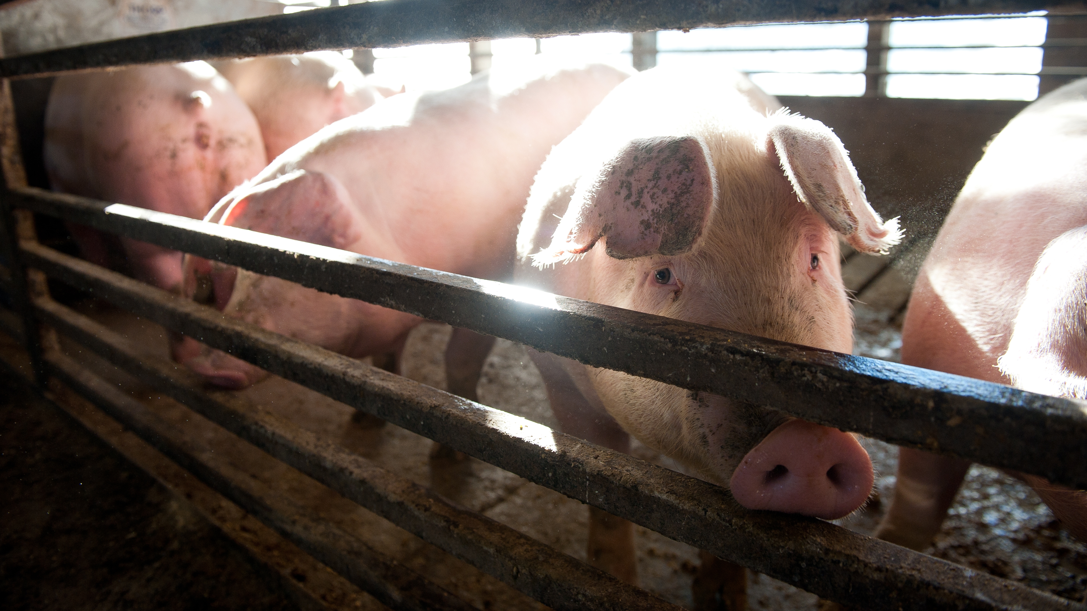
Porcine epidemic diarrhea
A 2-week-old pig was received for necropsy, from a farm with a history of diarrhea in piglets and vomiting in sows. The small intestine had transparent walls and watery contents. Histopathology revealed a marked shortening of intestinal villi, with necrosis/desquamation of epithelial cells and multifocal mild to moderate infiltrates of neutrophils and macrophages in the lamina propria, accompanied with occasional fibrin thrombi. Marked depletion of germinal centers was noted. Samples of intestine with fecal material were submitted to the Pennsylvania animal diagnostic laboratory in Harrisburg, PA for molecular testing, and Porcine epidemic diarrhea virus (a coronavirus) was detected. The infection with this virus can cause diarrhea in all ages of pigs, and can become a significant problem for pig farms if all ages are affected. In the USA, it was first described in 2013 and after that it has been reported in different states, including Virginia. Despite being a Coronavirus similar to Transmissible Gastroenteritis of pigs, samples do not cross react so it must be tested separately.
David Brown, DVM, RAHL Harrisonburg
Birds
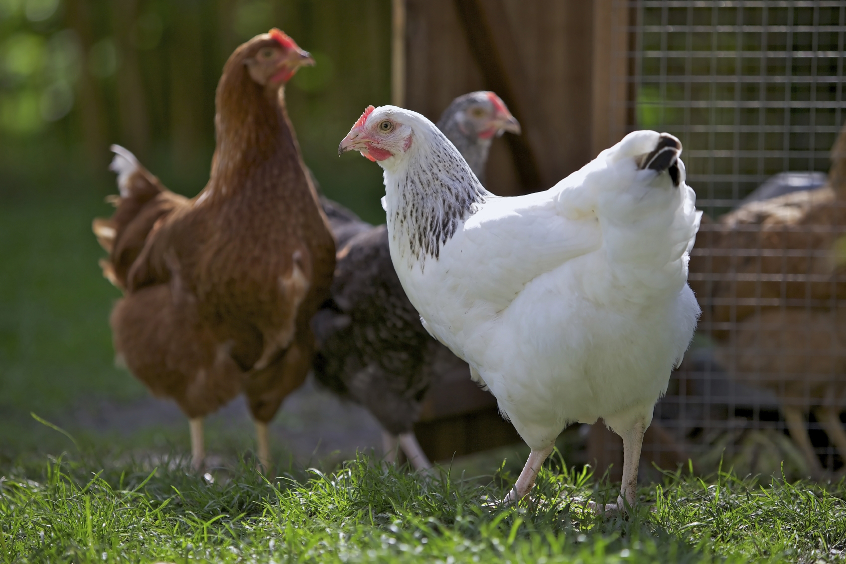
Avian Leukosis in a quail
A male quail was part of a collection seized from the breeder for suspected mismanagement to the point of cruelty. The bird had adequate fat stores as well as skeletal muscle. No obvious gross lesions were seen, but heart, liver, spleen, and intestines were all infiltrated with lymphoblasts. This is consistent with avian leukosis.
Phillip Sponenberg, DVM, PhD, Virginia Tech
Blackhead Disease in a Young Turkey Hen
Three 12-week old turkey hens were presented for necropsy. Gross necropsy revealed target lesions on the liver surface and throughout the parenchyma in 1 out of the 3 birds. Enlarged ceca with thickened walls and bilateral caseous cores were noted in the bird with target lesions. The other two birds had foamy, caramel colored cecal contents, but no evidence of thickened walls. Histopathology revealed transmural granulomatous typhlitis. Many macrophages and multinucleated giant cells have large round to crescent-shaped eosinophilic protozoa consistent with Histomonas meleagridis within the cytoplasm. Similar granulomas with protozoa were identified in the liver. Blackhead (or Histomoniasis) is a protozoal disease caused by Histomonas meleagridis. This protozoal is carried by the cecal worm (Heterakis gallinarum) as a host. It is commonly found to use earthworms as an intermediate host, and can be a persistent contaminant in the environment. This disease is particularly a risk when chickens and turkeys are housed together, because chickens can be asymptomatic carriers and shed the pathogen to highly susceptible turkeys.
Jessica Walters, DVM, PhD, DACPV, RAHL Harrisonburg
Companion/Exotic Animals

Wobbly hedgehog syndrome
A five-year-old female African pygmy hedgehog presented to a veterinarian for wobbliness/stumbling of several months’ duration and recent increase in respiratory effort. Significant physical exam findings, performed under light sedation, included muffled heart sounds, absent withdrawal reflexes in the right thoracic and pelvic limbs and dyspnea. The patient progressed to cardiopulmonary arrest while sedated and resuscitation attempt was unsuccessful. Post-mortem examination revealed multiple comorbidities including: 1. cardiomyopathy with resultant pulmonary congestion/edema, bi-cavitary effusion and chronic passive hepatic congestion; 2. bilaterally symmetrical spongy degeneration of the white matter throughout the brain most severe in the brainstem and cerebellum, consistent with wobbly hedgehog syndrome; and 3. biliary carcinoma of the liver. Wobbly hedgehog syndrome is an insidious, progressive neurologic disease of unknown cause affecting pet African pygmy hedgehogs.
Thomas Cecere, DVM, PhD, DACVP, Virginia Tech
Protozoal encephalitis in a puppy
An 11-week old, female Mastiff puppy presented to the VMCVM Necropsy service following a history of progressive paresis to full paralysis, with elective euthanasia due to poor quality of life. On gross examination, the hind legs were hyperextended and there was marked kyphosis of the lumbosacral spine, with marked, diffuse atrophy of the musculature of the hind legs. On histologic examination of the cerebrum, cerebellum, brain stem, cauda equina, and skeletal muscle from the left hindlimb, there were numerous, intracytoplasmic, protozoal cysts. Cysts measured up to 30 um in diameter and contained multiple, basophilic, 2um in diameter zoites. Associated lymphoplasmacytic inflammation was common. Cysts were also present within the sarcoplasm of myocytes. Definitive identification of the organism is pending, but initial differentials include Neospora caninum, Sarcocystis canis, Sarcocystis neurona, and Toxoplasma gondii. Based on location within the central nervous system as well as myocytes, Sarcocystis canis is of particular concern.
Vanessa Oakes, DVM, Virginia Tech
Laboratory News

ViTALS
ViTALS is excited to have become a new member of the National Animal Health Laboratory Network (NAHLN). Inclusion in the NAHLN allowed us to successfully compete for Farm Bill funding to contribute to chronic wasting disease (CWD) testing and improved results reporting in Virginia. It will also allow us to further support the VDACS laboratories in responding to high consequence veterinary diseases.
VDACS
The VDACS Wytheville Regional Animal Health Laboratory, in accordance with the AFS Blue Book requirements, has added bacteriology, virology, and parasitology testing for seven different diseases of cold-water fish raised in Virginia. Currently, the regulatory fish health inspections require testing for the following diseases: Furunculosis, Enteric Red mouth Disease, Bacterial Kidney Disease, Whirling Disease, Infectious Hematopoietic Necrosis Virus, Infectious Pancreatic Necrosis Virus, and Viral Hemorrhagic Septicemia Virus. Diagnostic testing for these same diseases is also available. Plans are being made to offer this testing for private health inspections, both in and out of state, as well as expanding the diseases tested in relation to other species.
Laboratory Locations
HARRISONBURG
261 Mt. Clinton Pike
Harrisonburg, VA 22802
540-209-9130
RAHLHarrisonburg@vdacs.virginia.gov
WARRENTON
272 Academy Hill Rd.
Warrenton, VA 20186
540-316-6543
RAHLWarrenton@vdacs.virginia.gov
LYNCHBURG
4832 Tyreeanna Rd.
Lynchburg, VA 24504
434-200-9988
RAHLLynchburg@vdacs.virginia.gov
WYTHEVILLE
250 Cassell Rd.
Wytheville, VA 24382
276-228-5501
RAHLWytheville@vdacs.virginia.gov
Virginia Tech Animal Laboratory Services
205 Duck Pond Drive
Blacksburg, VA 24061
540-231-7666
lcrvth@vt.edu